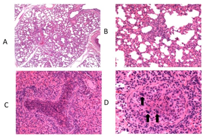
Figure 2.
Histological lesions observed in lung of calves after experimental infection with bovine RSV. Calves were challenged via aerosol with bovine RSV. On day 7 post-infection, samples of lung were collected for histological evaluation. A representative image from a single calf is shown. (A) Moderate interstitial thickening and wide interlobular septae due to edema. Original magnification, 4X. (B) Alveolar septae are thickened due to cellular infiltrates found to be macrophages, lymphocytes and lesser numbers of neutrophils when viewed at higher magnification. Original magnification, 20X. (C) Bronchioles are filled with neutrophils, sloughed epithelial cells, and necrotic cell debris. Original magnification, 20X. (D) There is partial to complete loss of bronchiolar epithelial cells with attenuation of remaining cells. In some bronchioles, epithelial cells form multinucleated syncytial cells (arrows). Original magnification, 40X. Figure adapted from: [31].

