
Figure 3.
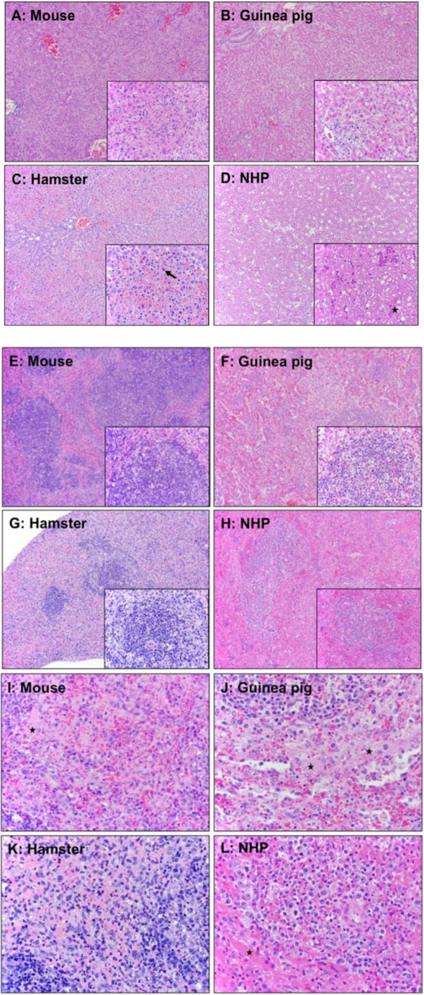
Comparison of pathology in mouse, guinea pig, hamster, and nonhuman primate. A Balb/c mouse and a Syrian hamster were infected IP with MA-EBOV; a Hartley strain of guinea pig was infected with GPA-EBOV; and a macaque was infected with wild-type EBOV. (A-D) Pathological changes in liver of different animal models. (A) Mouse: Multifocal, random hepatocellular degeneration and necrosis (10x and 40x inset). (B) Guinea pig: Diffuse, random hepatocellular degeneration and necrosis. Inflammatory cells are nearly absent (10x and 40x inset). (C–D) Hamsters: Liver. (C) Diffuse, midzonal hepatocellular degeneration, necrosis, and congestion. Inflammatory cells are nearly absent (10x). Solid arrow: prominent intracytoplasmic filovirus inclusion bodies in hepatocytes (40x). (D) Diffuse, random hepatocellular degeneration and necrosis (10x). Solid star: fibrin deposition (40x inset). (E-L) Pathological changes in spleen of different animal models. (E and I) Mouse: White and red pulp. White pulp (E); diffuse lymphoid necrosis and loss (10x and 40x inset). Red pulp (I); mild to moderate acute splenitis and small amounts of fibrin (solid star) (40x). (F and J) Guinea pig: White and red pulp. White pulp (F); multifocal lymphoid necrosis (10x and 40x inset). Red pulp (J); multifocal, mild to moderate acute splenitis with necrosis. Solid star: small amounts of fibrin at marginal zone (40x). (G and K) Hamster: White and red pulp. White pulp (G); diffuse lymphoid necrosis (10x and 40x inset). Red pulp (K); mild to moderate acute splenitis with monocytic degeneration and necrosis (40x). (H and L) NHP: White and red pulp. White pulp (H); diffuse lymphoid necrosis (10x and 20x inset). Red pulp (L); diffuse, moderate acute splenitis (40x). Solid star: fibrin.