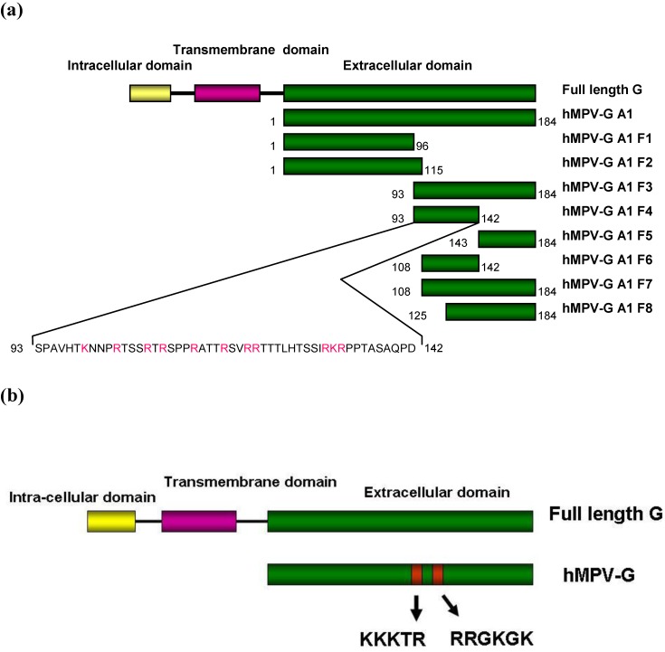
Figure 4.
A schematic diagram of recombinant hMPV G protein from (a) the A1 strain and the fragments produced and (b) the B2 strain. hMPV‑G A1 F1, F2, F3, F4, F5, F6, F7 and F8 indicate the 8 fragments of hMPV G A1 strain that were engineered. The sequence of the smallest fragment that binds to heparin (hMPV-G A1 F4) is shown with the positively charged residues in red. The red boxes represent the clusters of positively charged amino acids that are considered potential heparin binding sites.

