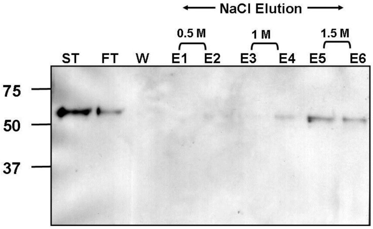
Figure 7.
Heparin agarose affinity chromotography of recombinant soluble hMPV-F protein. Start material (ST), flow through (FT), wash (W) and elution (E) fractions, with 0.5M NaCl (E1 & E2), 1M NaCl (E3 & E4) and 1.5M NaCl (E5 & E6), were analysed by 10% SDS-PAGE under reducing conditions and western blot analysis using anti-hMPV F antibody. Molecular weight markers are shown in kDa.

