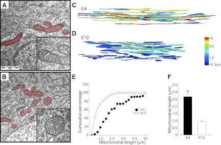
Figure 2.
Three-dimensional reconstruction of mitochondrial morphology in developing chick MNs. A, B) Ultrastructure of mitochondria in EM images shows intact cristae structure in E4 (A, inset) and E12 (B, inset) MNs. Mitochondria are pseudocolored in red. C, D) With the use of FIB, serial EM images were obtained; 3D reconstructed mitochondria on E4 (C) and E12 (D) are shown. Each mitochondrion is pseudocolored depending on its length. E, F) Direct measurement of mitochondrial length revealed a significant shortening of mitochondria between E4 and E12. *P < 0.05; Student's t test.

