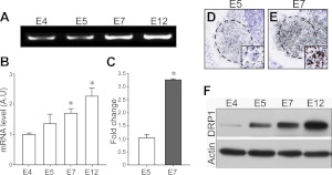
Figure 3.
Developmental expression patterns of DRP1. A–C) Changes in Drp1 mRNA expression in lumbar spinal cord (A) were quantified by RT-PCR (B) and real-time RT-PCR (C). *P < 0.05; 1-way ANOVA in Scheffe's multiple comparisons for post hoc comparisons; n = 5. D, E) In situ hybridization of Drp1 on E5 (D) and E7 (E) chick lumbar spinal cord. Dashed lines indicate lateral motor column (LMC). Insets: high-magnification view of single MNs in the LMC. F) Expression of Drp1 protein levels in developing spinal cord.

