Figure 4.
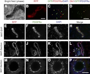
The aP2-lineage (RFP+) cells are associated with adipose vessels. A, B) Vessel from WAT SVP of aP2-Cre/Rosa26-tdTomato mice photographed under phase-contrast (A) and fluorescent (B) imaging. C) Cells cultured from SVP of aP2-Cre/Rosa26-EYFP mice coexpressed YFP (green) and PDGFRα (red). DAPI (blue) counterstain indicates nuclei. D) Coexpression of YFP (green) and Dlk1 (red). E–H) Whole-mount aP2-Cre/Rosa26-tdTomato SVP vessel was examined for expression of RFP (red) and PDGFRα (green). I–L) Sections of WAT labeled with RFP (red) and PDGFRα (green). M–P) Sections of BAT stained with RFP (red) and PDGFRα (green). Arrows indicate perivascular cells coexpressing RFP and PDGFRα. Scale bars = 100 μm.

