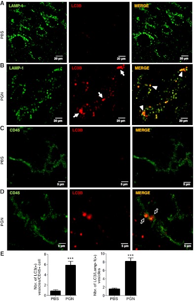
Figure 8.
PGN induces microglial cell autophagy in vivo. PBS (A, C) or PGN (B, D) was stereotaxically injected into the mouse CPU. After 24 h, 10-μm brain sections were stained with anti-LAMP-1 (green; A, B) or anti-CD45 (green; C, D) plus anti-LC3B (red) antibodies. Alexa Fluor 488 or Alexa Fluor 546 secondary antibodies were used. Slides were analyzed under a laser scanning confocal fluorescence microscope. Arrows indicate the presence of LC3B+ punctated cells. Arrowheads indicate colocalization of LC3B+ vesicles with LAMP-1. Bar graphs (E) represent means ± sd of number of LC3+ vesicles per CD45+ cell of 3 separate experiments. Number of LC3/Lamp-1 double-positive cells was obtained from 10 fields/slide, analyzing the next 3 sections without the needle artifact of 3 separate experiments. ***P < 0.001 vs. unstimulated cells.

