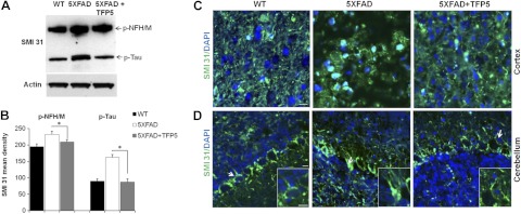
Figure 5.
TFP5 reduces hyperphosphorylation of neurofilament (p-NFH/M) in 5XFAD mice. A) Brain lysates from treated mutants show significant decrease in the expression of p-NFH/M, as detected by antibody SMI 31. Besides p-NFH/M, SMI 31 detects p-tau at 64 kDa. B) On quantification, both p-NFH/M and p-tau show significant decrease after TFP5 injections. Bars represent means ± se from 4 independent experiments. *P ≤ 0.05. C, D) Parasagittal brain sections incubated with SMI 31 antibody and analyzed under confocal microscope in the cortex (C) and cerebellum (D). Higher levels of p-NFH/M in 5XFAD mouse brain are reduced after TFP5 treatment. Arrows show the inset displaying aggregation of p-NFH/M in cell bodies, as observed in 5XFAD cerebellum. TFP5-treated brain cerebellum shows p-NFH/M staining in axons similar to WT mouse cerebellum. Scale bars = 20 μm.

