Figure 6.
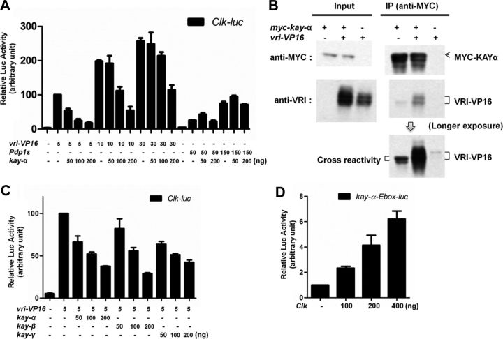
KAY-α interacts with and inhibits VRI. A, KAY-α blocks specifically VRI–VP16 activation of Clk promoter. HEK293 cells were transfected as indicated. Renilla luciferase was transfected to normalize transfection efficiency. Luciferase activity was measured 1 d after transfection. Relative luciferase activity with vri–VP16 was set to 100. VRI–VP16 activates the Clk promoter, as described previously (Cyran et al., 2003). The activation of the Clk promoter by VRI–VP16 was inhibited in a dose-dependent manner by KAY-α, but the activation of the Clk promoter by PDP1ε was unaffected. Error bars are SEM. B, KAY-α interacts with VRI–VP16 in HEK293 cells. HEK293 cells were transfected as indicated. Cell lysates were immunoprecipitated with anti-MYC antibody. Bound proteins were probed with anti-MYC and anti-VRI antibodies. VRI–VP16 was coimmunoprecipitated with MYC–KAY-α. C, KAY-β and KAY-γ can also repress the activation of the Clk promoter by VRI–VP16. HEK293 cells were transfected as indicated. A Renilla-expressing vector was cotransfected to normalize transfection efficiency. The normalized luciferase activity with vri–VP16 was set to 100 on the graph. Error bars are SEM. D, CLK can activate the kay-α promoter. A ∼300 bp kay-α promoter fragment containing an E-box was cloned in the pGL3 vector to generate kay–α-Ebox–luc. It can be activated by CLK. The normalized luciferase activity without Clk was set to 1 on the graph. Error bars are SEM.

