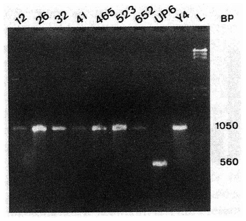
Fig. 2.

Analysis of the leukotoxin status of A. actinomycetemcomitans strains by PCR amplification of the lkt promoter region. This photograph shows the migration in an agarose gel of amplicons resulting from PCR amplification of the lkt promoter region, as described in materials and methods. The numbers at the top of the gel represent the different A. actinomycetemcomitans strains. The letter L corresponds to the Lambda standard (λ HindIII). Numbers at the right represent the size of the amplicon for nonleukotoxic (1050 bp) and leukotoxic (560 bp) strains.
