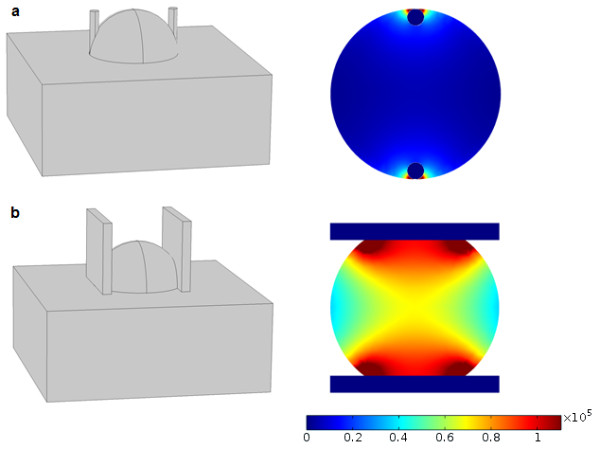
Figure 1.

Electric field distribution for plate electrodes in a cutaneous tumor electroporation (a) and parallel needle electrodes in cutaneous tumor electroporation (b). Geometry is shown on the left hand side and resulting field strength on the right. Tumor diameter in both instances is 2 cm, electrode thickness 0.2 cm and electrode distance 1.6 cm. The applied voltage was 1300 V. Resulting field strength on the color scale is in volts per meter.
