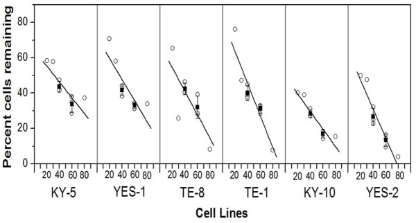
Figure 2.
Effects of curcumin on six ESCC cell lines. At 60 μM curcumin, YES-2 and KY-10 were more sensitive than KY-5, YES-1, TE-8, and TE-1 (p<0.05, ANOVA, F=16.44, p<0.0001). Cell loss was measured by a standard crystal violet assay after incubation with 20 to 80 μM curcumin for 30 hrs. Each point represents the effect of one curcumin dosage (20, 30, 40, 60 or 80 μM). For 40 and 60 μM the error bar indicates mean (±SD) of three experiments.

