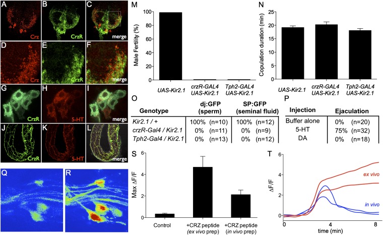
Fig. 3.
Corazonin receptor-GAL4 neurons innervate reproductive organs and control sperm transfer. (A–C) Confocal image of crzR-GAL4; UAS-mCD8-GFP male AG stained with anti-corazonin (red). (D–F) Higher-magnification view (boxed region in C) of the AG showing the intermingling of corazonin and crzR-GAL4 terminals. (G–L) crzR-GAL4; UAS-mCD8-GFP male labeled with antibodies to GFP (green) and 5-HT (red). (G–I) High-magnification view of sexually dimorphic abdominal ganglia neurons expressing both 5-HT and crzR-GAL4. (J and K) Overlapping projections innervating the accessory glands. (M) Fertility of the indicated genotypes after mating with wild-type virgins. n ≥ 30 per genotype. (N) Copulation duration of crzR-GAL4; UAS-Kir2.1 (20 ± 2 min, n = 15), Tph2-GAL4/Kir2.1 (18 ± 2 min, n = 16), and Kir2.1/+ (19 ± 2 min, n = 14) males. (O) Assay for transfer of sperm (dj:GFP) and seminal fluid (SP:GFP) to females during mating. (P) Individual male flies were injected with 5-HT, dopamine, or buffer and observed for ejaculation (Materials and Methods). (Q and R) Z-projection image of a crzR-Gal4; UAS-GCaMP5G abdominal ganglia before (Q) and after (R) Crz bath application. (S) Peak ΔF/F values (n = 3–5 cells from at least three crzR-Gal4; UAS-GCaMP5G flies for each condition) following bath application of buffer (control) or the Crz peptide dissolved in buffer. Crz was applied either to an isolated nerve cord (ex vivo prep) or an intact fly with the AG exposed (in vivo prep). (T) Example ΔF/F responses from ex vivo (red) and in vivo (blue) preparations. Traces have been aligned to the rise of the response. Crz peptide (or buffer alone) was added before time = 0 and washed out 300 seconds later.

