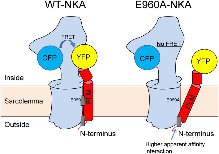
Fig. 7.
Working model of NKA–PLM complex with attached CFP and YFP. Left shows WT–NKA (blue) and WT–PLM (red). FRET occurs between CFP and YFP and the complex is held together by two binding regions (gray rectangles): E960–F28 functional interaction region and a second region. Right shows a different NKA–PLM conformation in the E960A mutant (or F28A mutant). CFP and YFP are not sufficiently close for appreciable FRET, but the complex is still held together by a higher apparent affinity interaction region near the external membrane surface.

