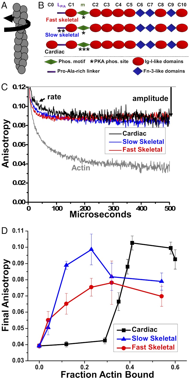
Fig. 1.
(A) Actin filament twisting motions in the μs time range are detected by TPA. (B) Domain organization of MyBP-C isoforms. Domains listed from N-terminal C0 to C-terminal C10, including the Pro/Ala rich linker (LP/A) and phosphorylation motif (m). (C) Effect of 1 μM fast skeletal (red), 1 μM slow skeletal (blue) or 4 μM cMyBP-C (black) on the TPA decay of ErIA-actin (gray). (D) Dependence of final anisotropy on actin binding.

