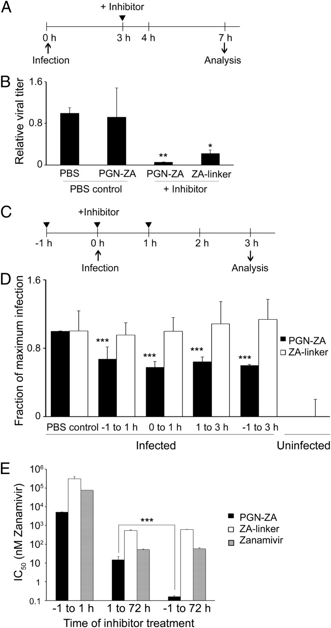
Fig. 2.
PGN-ZA synergistically inhibits early and late steps of influenza virus infection. (A) Experimental design to detect the release of newly synthesized viruses from infected cells. (B) MDCK cells were inoculated with WSN virus in a synchronized infection, and unbound virus was then removed. The inhibitors were added 3 hpi. The supernatant was collected at 7 hpi., serially diluted, and titrated using the plaque assay. As a control for any remaining inhibitor in the plaque assay, some PBS control samples were spiked with the same concentration of PGN-ZA and titrated in parallel with the original PBS controls. (C) Scheme of time-of-addition experiment to assay inhibition in the early phase of virus infection in a single replication cycle assay. (D) MDCK cells were infected with WSN, and the inhibitors were added in at −1, 0, or 1 hpi. The cells were trypsinized, fixed at 3 hpi., stained for intracellular viral NP and M1, and analyzed by flow cytometry. The gating for virus-infected cells was drawn based on the expression level of a mock-infected control, and the fraction of the cells infected was normalized to the untreated, infected sample. (E) Synergistic inhibition of early and late steps of influenza infection by PGN-ZA. MDCK cells were infected with A/Turkey/MN/80, and the inhibitors were added either −1 to 1 hpi, 1 to 72 hpi, or −1 to 72 hpi. The bars represent IC50 values (i.e., the concentration of inhibitor reducing the plaque number in untreated controls by half). For the samples in the −1 to 1 h condition, the IC50s are higher than the values indicated, as we did not observe any significant plaque number reduction at this concentration. Error bars in B, D, and E represent SEM from three to five independent experiments. *P < 0.05, **P < 0.01, ***P < 0.001.

