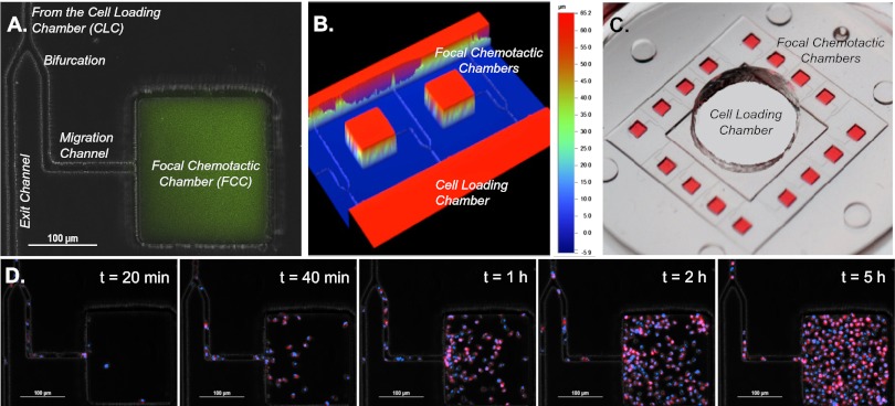
Fig. 2.
Characterization of microfluidic postdiapedesis inflammation model. (A) Chemoattractant (green) is primed into the device and a gradient is formed along the migration channel toward the FCC. (B) Profilometer image of two adjacent FCCs illustrate distinct heights of FCCs and migration channels. (C) Large-scale model of device. (Magnification, 5×.) Sixteen FCCs (red) surround each CLC. After washing, the chemoattractant only remains in the FCC, and a linear gradient is formed along the migration channel. (D) Double-stained (blue, nucleus; red, membrane) neutrophils begin migrating along the LTB4 gradient after 20 min and fill the chamber by 5 h (Movie S1).

