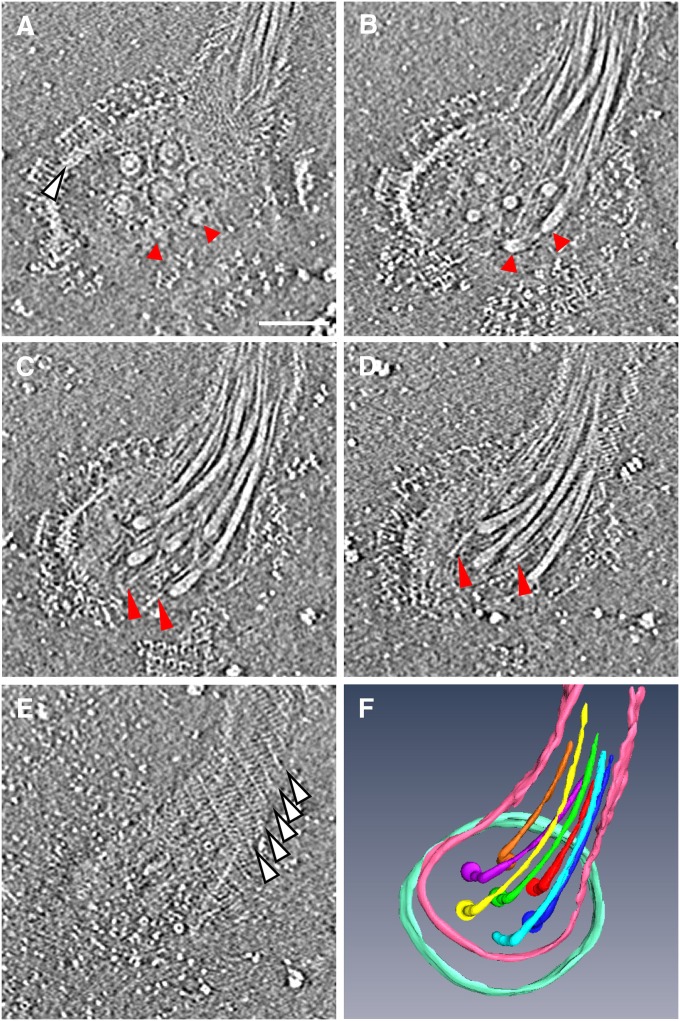
Fig. 2.
Architecture of the flagellar apparatus detached from the cell body. (A–E) Five selected slices of a tomogram from lower to upper positions. The slice distance is 18.9 nm from A to B, and 9.44 nm from B to C, C to D, and D to E. The flagellar apparatus is viewed from outside of the cell. (Scale bar, 100 nm.) (A) Slice showing seven flagellar basal bodies in the hexagonal array. Each basal body shows the L and P ring and the rod within the rings. The upper five are clear, but the bottom two, indicated by red arrowheads, are only partially visible in this slice. The edge of the base platform is indicated by a white arrowhead. (B) Slice showing the proximal end of flagella in the hexagonal array. Two arrowheads indicate the ends from which two flagella are extend upward. (C and D) Slices showing the flagella and fibrils. Fibrils are indicated by red arrowheads. (E) Slice showing the upper surface of the sheath. White arrowheads indicate the helical lines shown in Fig. 1E. (F) Solid surface rendered by 3D segmentation of the seven flagella, part of the sheath, and the edge of the base platform. The tomogram was reconstructed from a tilt-series of cryoEM images from −50° to 70° with an increment of 2°.

