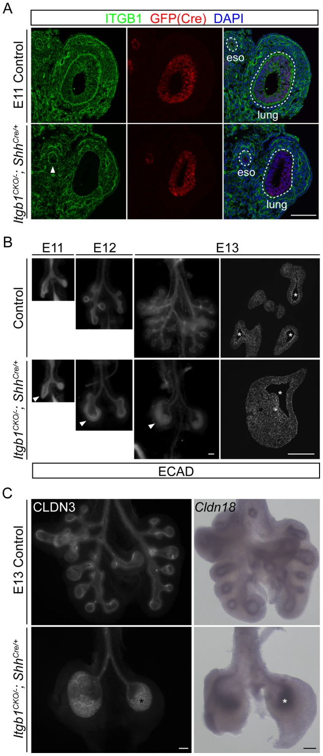
Figure 1. Epithelial inactivation of Itgb1 inhibits branching and leads to a multilayer epithelium during lung development.

(A) Section immunostaining showing complete loss of ITGB1 specifically in the lung epithelial cells, but not the surrounding mesenchymal cells as early as embryonic day 11 (E11) in the Itgb1CKO/−; ShhCre/+ mutant. Sections were co-stained for GFP to visualize cells expressing the GFP-CRE fusion protein under the control of the Shh promoter, and nuclei were counter-stained with 4′,6-diamidino-2-phenylindole (DAPI). The arrowhead indicates partial loss of ITGB1 in the ventral half of the esophagus epithelium in the Itgb1CKO/−; ShhCre/+ mutant. The dashed lines demarcate the basal side of the lung and esophagus (eso) epithelia. Scale bar, 100 um. (B) Whole-mount (left three columns) and section (right column) immunolocalization of E-Cadherin (ECAD) showing the inhibition of branching and progressive formation of a multilayer lung epithelium (arrowheads) at sequential embryonic days (E11, E12, E13) in the Itgb1CKO/−; ShhCre/+ mutant. Because of the three-dimensional structure of the lung, the epithelium can artificially appear multilayered in tangential sections through the epithelium. The number of epithelial layers can only be accurately assessed in regions of sections where the lumenal space (asterisks) is visible; note that there are more than 10 cell layers in the E13 mutant lung. Scale bar, 100 um. (C) Whole-mount immunostaining (left panels) and in situ hybridization (right panels) of E13 lungs showing that cells (asterisks) of the multilayer epithelium in the Itgb1CKO/−; ShhCre/+ mutant lung maintain expression of epithelial markers CLDN3 (left panels) and Cldn18 (right panels). Scale bar, 100 um.
