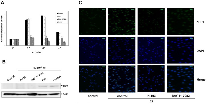
Figure 3. E2 treatment regulates δEF1 expression via PI3K and NF-κB pathways.
A. MCF-7 cells were pre-incubated with PKI (5 µg/µl), BAY 11-7082 (10 µM), or PI-103 (10 µM) for 0.5 h followed by treatment with 10−9 M E2. The expression of δEF1 mRNA was determined at the indicated time points following treatment using Q-PCR. GAPDH was used to normalize the δEF1 level. * indicates p<0.05 in unpaired Student’s t-test compared with control. ** indicates p<0.01 in unpaired Student’s t-test compared with control. The data represent three independent experiments. B. MCF-7 cells were pre-incubated with PKI, BAY 11-7082, or PI-103 for 0.5 h followed by treatment with 10−9 M E2. The expression of δEF1 protein was determined at 24 h following treatment using Western Blot. Actin was used to normalize the δEF1 level. C. MCF-7 cells were pre-incubated with PKI, BAY 11-7082, or PI-103 for 0.5 h followed by treatment with 10−9 M E2. The expression of δEF1 protein was determined at 24 h following treatment using immunofluorescence. Scale bars, 50 µm.

