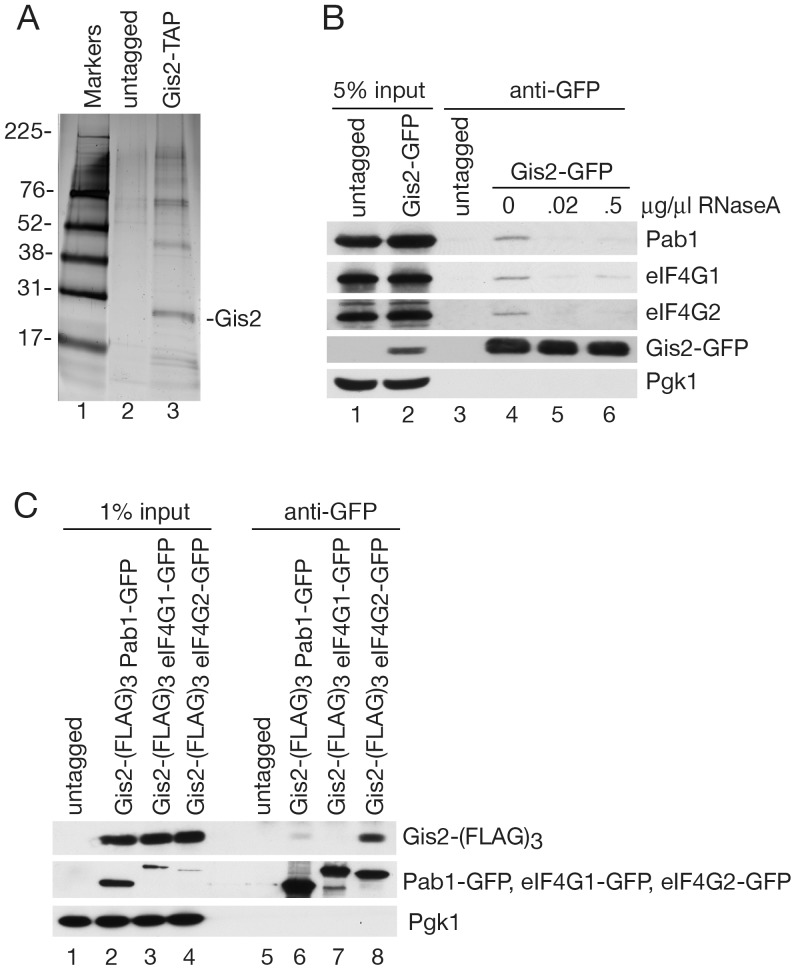
Figure 1. Gis2 associates with proteins involved in translation initiation.
(A) After tandem affinity purification, eluates from an untagged strain and a strain expressing Gis2-TAP were fractionated in a SDS-polyacrylamide gel and proteins visualized by silver staining. Lane 1, molecular size markers. Sizes are in kDa. (B) Lysates of an untagged strain and a strain expressing Gis2-GFP were subjected to immunoprecipitation with anti-GFP antibodies. Prior to immunoprecipitation, Gis2-GFP lysates were incubated with the indicated amounts of RNase A. Proteins in immunoprecipitates were detected by Western blotting with antibodies against Pab1, eIF4G1, and eIF4G2. The efficiency of immunoprecipitation was determined by re-probing with anti-GFP. As a negative control, the blot was reprobed to detect Pgk1. (C) Lysates of untagged and Gis2-(FLAG)3 strains expressing Pab1-GFP, eIF4G1-GFP or eIF4G2-GFP were subjected to immunoprecipitation with anti-GFP antibodies. After Western blotting, Gis2-(FLAG)3 was detected with anti-FLAG antibodies. To examine immunoprecipitation efficiency, Pab1-GFP, eIF4G1-GFP and eIF4G2-GFP were detected with anti-GFP antibodies. Pgk1 was detected as a negative control.

