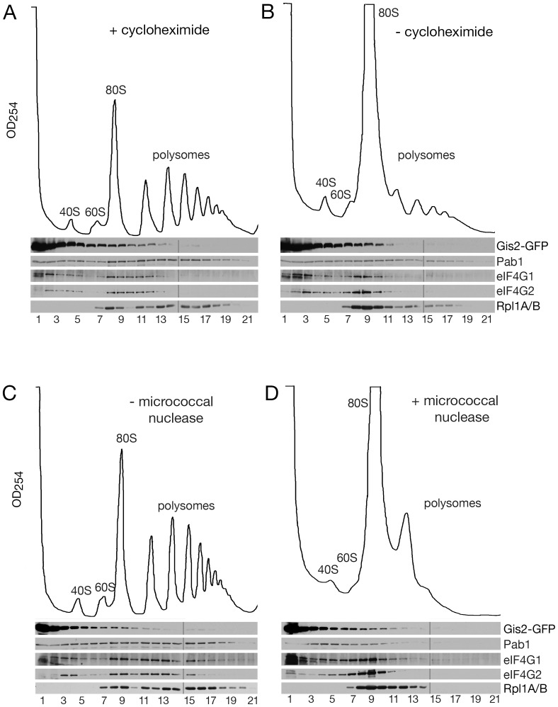
Figure 2. A small fraction of Gis2 sediments with polyribosomes.
(A and B) GIS2-GFP cell lysates were prepared in the presence (A) or absence (B) of cycloheximide and fractionated in 15–50% sucrose gradients. Fractions were collected while monitoring OD254. Proteins were subjected to Western blotting to detect Gis2-GFP, Pab1, eIF4G1, eIF4G2 and ribosomal proteins L1A and L1B. (C and D) GIS2-GFP cell lysates prepared in the presence of cycloheximide were either untreated (C) or incubated with 5 U/µl micrococcal nuclease (D) prior to sedimentation. Fractions from each gradient were analyzed in two gels as indicated by the lines.

