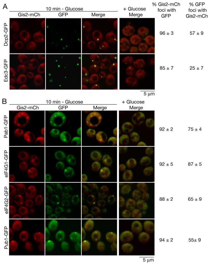
Figure 3. Gis2 accumulates in P-bodies and stress granules during glucose depletion.
(A and B) Yeast strains expressing chromosomal Gis2-mCh and (A) the P-body markers Dcp2-GFP and Edc3-GFP or (B) the stress granule markers Pab1-GFP, eIF4G1-GFP, eIF4G2-GFP and Pub1-GFP were grown in glucose-containing media, then resuspended in fresh media that either lacked or contained glucose. After 10 minutes, cells were observed using confocal microscopy. In glucose media (right column), no Gis2-mCh foci were observed; thus only the merged panels are shown. Bars, 5 µm.

