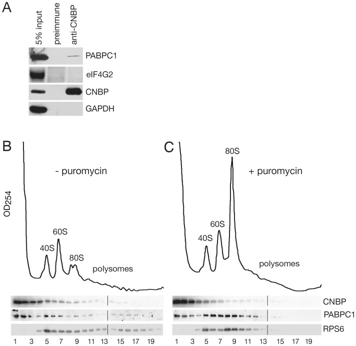
Figure 6. Some CNBP associates with PABPC1 and sediments with translating ribosomes.
(A) HeLa cell lysates were subjected to immunoprecipitation with anti-CNBP antibodies. Proteins in immunoprecipitates were subjected to Western blotting to detect the poly(A) binding protein PABPC1 and eIF4G2. To assess the efficiency of immunoprecipitation, the level of CNBP in the immunoprecipitate was also determined. As a negative control, the blot was reprobed to detect GAPDH. (B and C) HeLa cells were either untreated (B) or incubated with puromycin for 20 minutes (C) prior to harvesting in cycloheximide. Lysates were sedimented in 15–50% sucrose gradients and fractions collected while monitoring OD254. Proteins were subjected to Western blotting to detect CNBP, PABP1C and ribosomal protein RPS6.

