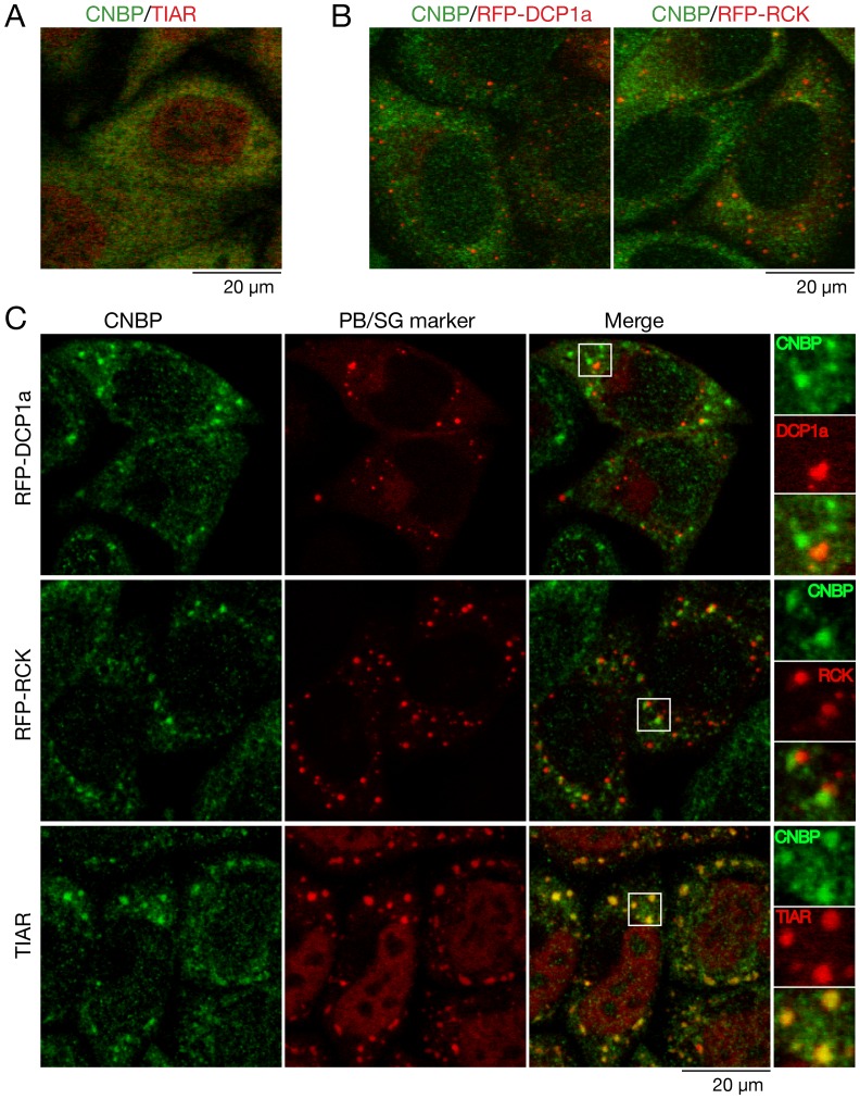
Figure 7. CNBP accumulates in stress granules during arsenite treatment of HeLa cells.
(A) HeLa cells were subjected to immunofluorencence to detect CNBP (green) and the stress granule marker TIAR (red). A merged image is shown. Bar, 20 µm. (B) HeLa cells transfected with plasmids expressing RFP-DCP1a (left) or RFP-RCK (right) were subjected to immunofluorescence with anti-CNBP antibodies. Merged images are shown. Bar, 20 µm. (C) To induce P-bodies and stress granules, untransfected cells and cells expressing RFP-DCP1a or RFP-RCK were incubated with 500 µM arsenite for 30 minutes. Following immunofluorescence to detect CNBP (top and middle panels) or both CNBP and TIAR (bottom panel), cells were examined using confocal microscopy. Bar, 20 µm. The rightmost panels show enlarged images of the boxed areas.

