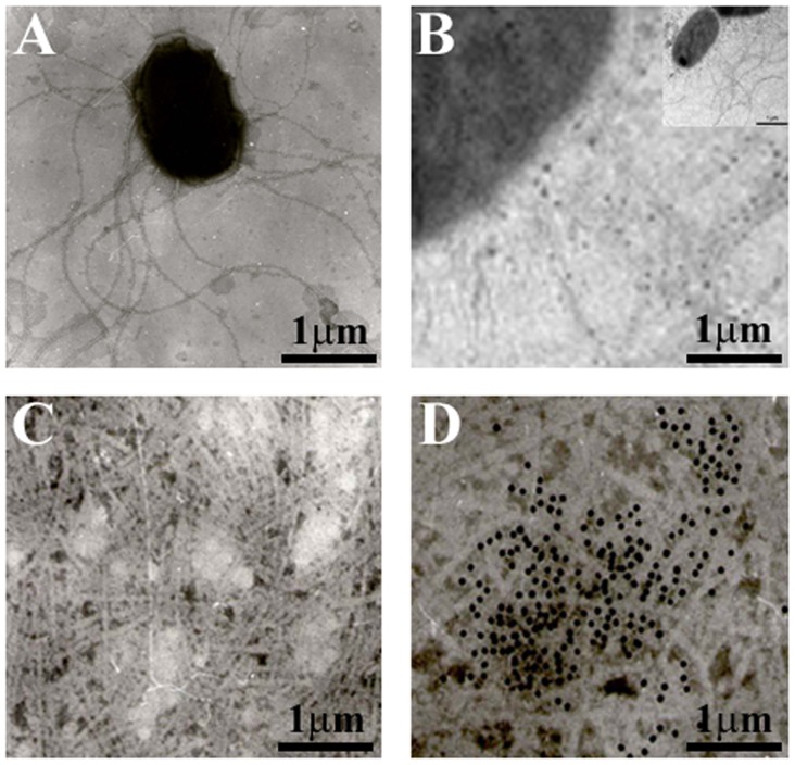
Figure 1. Flagella identification by transmission electron microscopy.
(A) C. sakazakii ATCC BAA-894 strain shows flagellar structures protruding from the bacteria after growth on TSA agar at 37°C. (B) Immunogold-labeling of flagella produced by C. sakazakii on TSA agar using anti-flagella antibody. The inset shows the micrograph of the whole bacteria using immunogold-labeling obtained by TEM. (C) Purified flagella. (D) Immunogold-labeling of purified flagella. Samples were negatively stained and electron micrographs were taken at a magnification of 19,000x.

