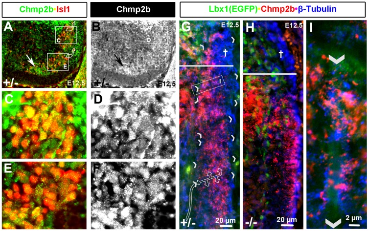
Figure 8. Chmp2b Protein in Soma and Dendrites of Motor Neurons.
Chmp2b and Isl1 are co-expressed in motor columns of the ventral horn. (A, C, E) Isl1 primary antibody was detected with Cy3 (red). Chmp2b antibody was detected with Cy5 (infrared) and is shown in green. (B, D, F) Greyscale images of the green channel of images shown in A, C, and E, respectively. Chmp2b staining within soma can be distinguished from staining of projections between soma. The most sensitive secondary antibody (Cy3) and relatively long image acquisition times are required to detect Chmp2b signal in the motor columns. (G, H) Comparison of the VLF of heterozygotes and mutants stained with Chmp2b (red; Cy3) and tubulin (blue; Cy5). Radial breaks in the tubulin stains of heterozygotes (indicated by pairs of arrowheads) may represent the endfeet of radial glial cells (see inset I) that dendrites of motor neurons have been shown to follow into the VLF during early synaptogenesis (diagrammed by flow arrow). Note the loss of tubulin staining in the LF (†) and in the VLF below the sulcus limitans (horizontal line). Chmp2b staining in the VLF is present but distributed differently in mutants. (I) High magnification image of a putative radial glial endfoot that can be seen by background stain in the green channel. Note that Chmp2b and tubulin staining do not colocalize but appear to associate closely on the surface of the endfoot.

