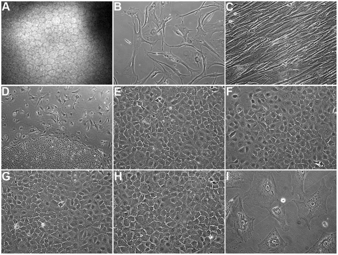
Figure 1. Morphologic study of corneal endothelial primary cells.
(A) In vivo confocal microscopy of human corneal endothelium demonstrating the typical hexagonal cell morphology. (B) Phase-contrast micrograph of non-dividing primary cells from a 21-year-old donor (21M) at passage 9, displaying typical signs of senescence. 200x. (C) Phase-contrast micrograph of primary cells from a 63-year-old donor that underwent endothelial-mesenchymal transformation at passage 1. Note that the cells lost the typical corneal endothelial morphology and appear fibroblast-like. 200x. (D) Phase-contrast micrograph showing 2 morphologically distinct subpopulations of cells in the 21M primary culture. The uniform cells growing in colony-like structures were designated HCEnC-21. 40x. (E-H) Phase-contrast micrographs of HCEnC-21 at passages 24 (E) and 46 (F) as well as telomerase-transduced HCEnC-21 (HCEnC-21T) at passages 25 (G) and 50 (H). Importantly, HCEnC-21 and HCEnC-21T cells grew in contact-inhibited monolayers displaying the typical hexagonal cell morphology seen in vivo. 200x. (I) Phase-contrast micrograph of telomerase-transduced 21M (21M+hTERT) at passage 7 showing a senescent phenotype. 200x.

