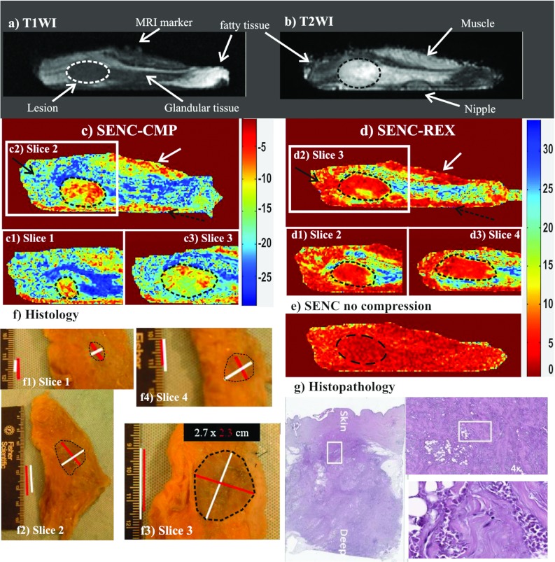
Figure 9.
Demonstration of breast SENC MRI method on an ex vivo breast sample with a mass determined to invasive ductal carcinoma (unknown before imaging). Representative SENC breast images of (a) T1-weighted image, (b) T2-weighted image with fat suppression, (c) breast SENC-CMP image of the breast tissue, (d) SENC-REX image of the breast tissue, and (e) breast SENC MRI image with no compression shows homogeneous strain throughout the breast. Indicating no residual strain within the breast tissue without any compression applied. Dotted ROI shows the breast lesion in all image locations. (c1)–(c3) and (d1)–(d3) Breast SENC-CMP and SENC-REX images show different slices containing the tumor. White arrows point to muscle, while black and dotted black arrows point to image artifacts. (f) and (g) Histological results clearly demonstrate the breast lesion is invasive ductal carcinoma. The histological images show the gross pathology and the microphotograph of the H&E stained section.

