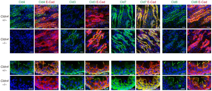
Figure 2. Compensatory accumulation of other Clds at TJs in the nephrons and urothelium of Cldn4 −/− mice.
Renal medullary regions (upper) and ureters (lower) of Cldn4+/− mice and Cldn4−/− mice (2 months old) with no evidence of hydronephrosis were three-color immunostained with anti-Cld4, anti-Cld3, anti-Cld7, or anti-Cld8 (green), along with anti-E-cadherin (E-cad) (red) and DAPI (blue). Bars, 50 µm (upper) and 20 µm (lower). Note the increased signal of Cld3 with diminished Cld8 signal at the cell-cell adhesion sites in renal medullary regions and increased Cld7 signal in ureter of Cldn4−/− mice.

