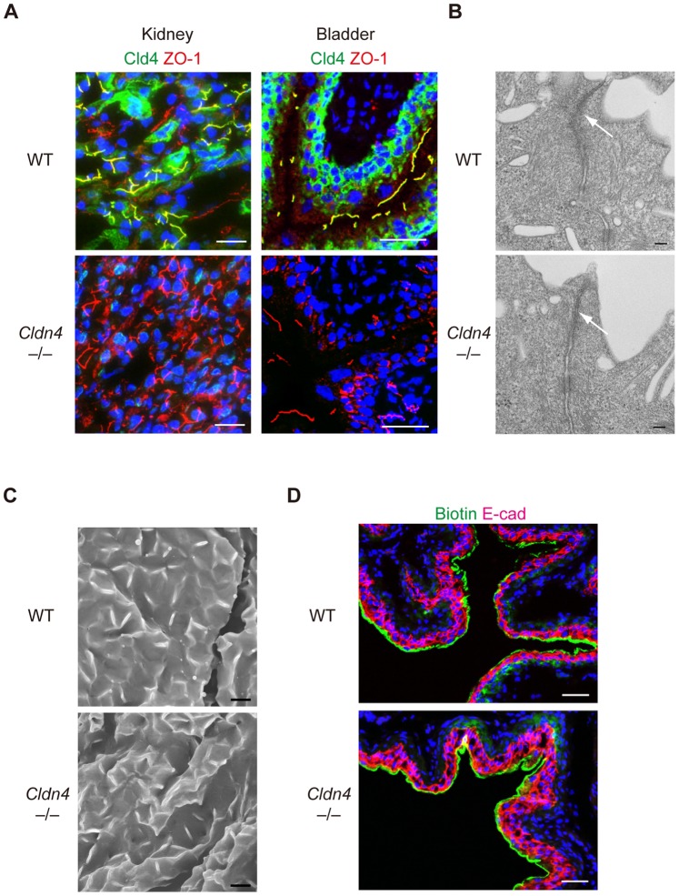
Figure 3. Preserved TJs and barrier function of TJs of Cldn4 −/− mice.
(A) Kidneys and bladders of WT and Cldn4−/− mice were immunostained with anti-Cld4 (green), anti-ZO-1 (red), and DAPI (blue). Bars, 50 µm. (B) Urothelium of ureters of WT and Cldn4−/− mice were subjected to ultrathin transmission electron microscopy. Bars, 100 nm. Arrows indicate TJs. (C) Urothelium of bladders of WT and Cldn4−/− mice were subjected to scanning electron microscopy. Bars, 5 µm. (D) WT and Cldn4−/− mice under anesthesia were injected with sulfo-NHS-SS-biotin into bladders, and 20 minutes later the bladders were dissected and 3-color immunostained with streptavidin (green) and E-cadherin (E-cad)(red) and DAPI (blue). Bars, 50 µm.

