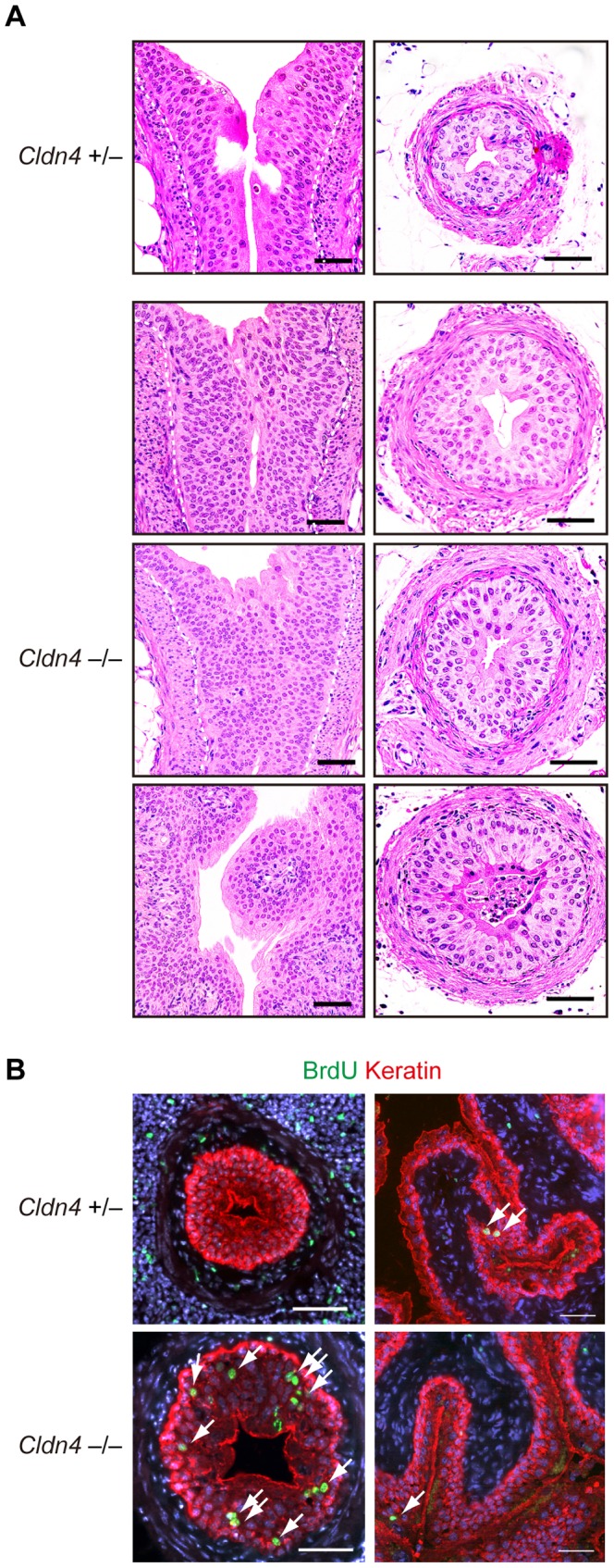
Figure 6. Urothelial hyperplasia with increased BrdU incorporation in Cldn4 −/− mice.

(A) Sections of the uretero-pelvic junction regions (left panels) and lower part of the ureters (right panels) of Cldn4 +/− and Cldn4 −/− littermate mice (8–19 month old) with no evidence of hydronephrosis were stained with hematoxylin and eosin. Dotted lines indicate the urothelial basal layers. Note the increased urothelial cell numbers and layers. Bars, 50 µm. (B) Cldn4 +/− and Cldn4 −/− littermate mice (12 months old) were injected with BrdU (1 mg/animal) intraperitoneally for 4 consecutive days, and the sections of the ureters (left) and bladders (right) were three-color immunostained with anti-BrdU (green, arrows), anti-keratin (red), and DAPI (blue). Bars, 50 µm.
