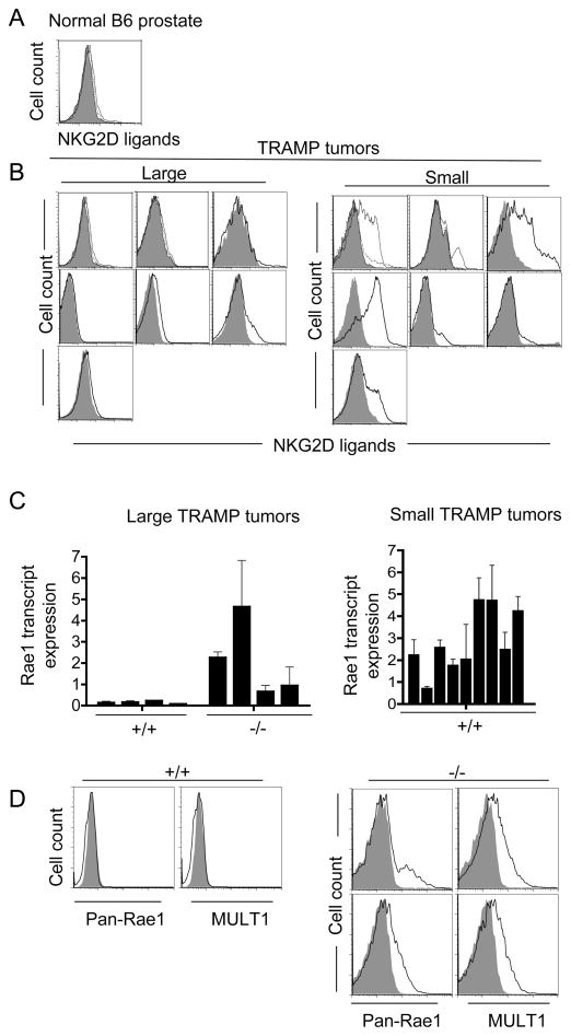
Figure 3. Expression of NKG2D ligands by prostate carcinomas ex vivo.
(A, B) Reduced cell surface expression of NKG2D ligands on large, early-arising B6-TRAMP prostate tumors as compared to smaller, late-arising ones. Freshly dissociated prostate tissue from a representative non-transgenic Klrk1+/+ B6 mouse (A) and sets of large and smaller tumors isolated from Klrk1+/+ B6-TRAMP mice (B) were compared. Labeled NKG2D tetramers (plain line) were used to detect all NKG2D ligands and streptavidin-PE served as a negative control (shaded histogram). In a few cases, the specificity of staining was proved by inhibition with unlabeled NKG2D tetramers (dotted line). (C) Reduced amounts of Rae1 transcripts in large (>2.7 g) Klrk1+/+ TRAMP tumors as compared to large Klrk1−/− TRAMP tumors (p=0.0286 with the Mann-Whitney test) (left panel) or smaller Klrk1+/+ TRAMP tumors (right panel). Values determined by quantitative RT-PCR with primers that detect all Rae1 isoforms were normalized to HPRT transcript amounts, and these data were normalized to the amount in nontransgenic B6 prostates (average of 5 independent B6 mice). Graphs show the mean ± SD of 2–6 separate qPCR assays for each sample. (D) Cell surface staining of Rae1 and MULT1 (solid line) on two dissociated large Klrk1−/− TRAMP tumors and a large tumor from an Klrk1+/+ TRAMP littermate. Isotype control stains are shown as shaded histograms. Gated CD45-negative (non-hematopoietic), PI-negative (live) cells were examined.

