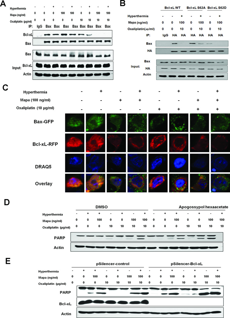
Figure 5. Role of Bcl-xL in apoptosis: The dissociation of Bcl-xL from Bax in oxaliplatin/Mapa/hyperthermia treated CX-1 cells.
(A) Cells were treated and cell lysates were immunoprecipitated with anti-Bax antibody or mock antibody (IgG) and immunoblotted with anti-Bcl-xL or anti-Bax antibody (upper panels). The presence of Bcl-xL and Bax in the lysates was verified by immunoblotting (lower panels). (B) Transfectants with Bcl-xL-WT, BclxL- S62A, or Bcl-xL S62D were treated with oxaliplatin/Mapa/hyperthermia and were immunoprecipitated with anti-HA antibody or IgG and immunoblotted with anti-Bax or anti-HA antibody (upper panels). The presence of HA, Bax and actin in the lysates was examined (lower panels). (C) Cells were cotransfected with pBax-GFP and pBcl-xL-RFP plasmid, and 24 h later treated with oxaliplatin/Mapa/hyperthermia. Cellular DNA was stained with DRAQ5. Localization of Bax-GFP and Bcl-xL-RFP was examined by confocal microscope. (D) Cells were pretreated with apogossypol hexaacetate followed by oxaliplatin/Mapa/hyperthermia. PARP cleavage was detected. (E) Transfectants with pSilencer-control or pSilencer-Bcl-xL were treated with oxaliplatin/Mapa/hyperthermia and immunoblotted with anti-PARP or anti-Bcl-xL antibody. Actin was shown as an internal standard.

