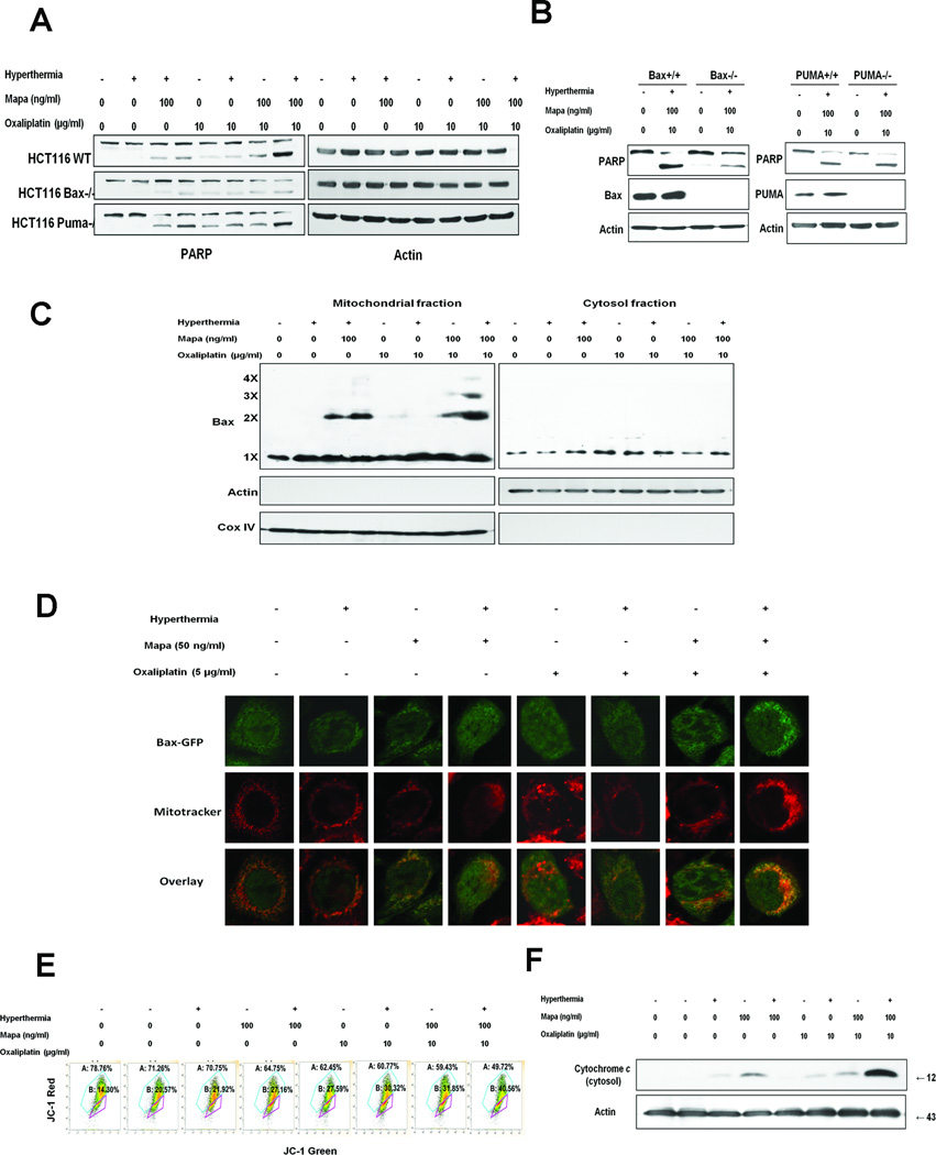
Figure 6. Bax oligomerization, localization to the mitochondria and subsequent cytochrome c release in Mapa/oxaliplatin/hyperthermia.
(A) Human colon carcinoma parental HCT116 (HCT116 WT), Bax knockout HCT116 Bax−/−and PUMA knockout HCT116 PUMA−/− cells were treated with oxaliplatin/Mapa/hyperthermia and immunoblotted with anti-PARP antibody. B HCT116 Bax+/+HCT116 Bax−/−HCT116 PUMA+/+ and HCT116 PUMA−/− cells were treated with oxaliplatin/Mapa/hyperthermia and immunoblotted with anti-PARP, anti-Bax, or anti-PUMA antibody. (C) CX-1 cells were treated with oxaliplatin/Mapa/hyperthermia. Mitochondrial and cytosolic fractions were isolated and were cross-linked with dithiobis and subjected to immunoblotting with anti-Bax antibody. Bax monomer (1X) and multimers (2X, 3X, and 4X) are indicated. Actin was used as a cytosolic marker and COX IV as a mitochondrial marker. (D) CX-1 cells were transfected with pBax-GFP plasmid. After 24 h incubation, cells were treated with oxaliplatin/Mapa/hyperthermia. Mitochondria were stained red with MitoTracker. Localization of Bax-GFP was examined by confocal microscope. (E) CX-1 cells were treated with oxaliplatin/Mapa/hyperthermia and stained with JC-1 and analyzed by flow cytometry. (F) CX-1 cells were treated with oxaliplatin/Mapa/hyperthermia. Cytochrome c release into cytosol was determined by immunoblotting for cytochrome c in the cytosolic fraction. Actin was used to confirm the equal amount of proteins loaded.

