
Figure 2. tEGF protects differentiating osteogenic cells from death signals.
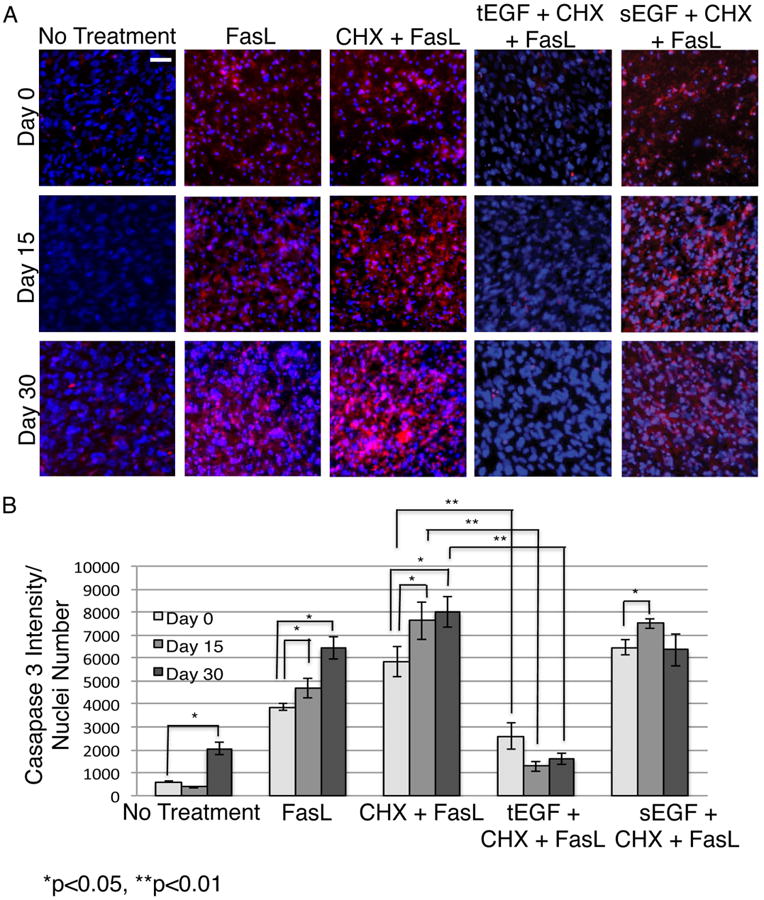
Fluorochrome inhibitor of caspase assay (FLICA) stained imhMSC (Magnification 10×, Scale bar 50μm) grown on mock/tEGF surfaces under osteogenic conditions, after 8 hours of treatment with cytokines on Day 0/ Day 15/ Day 30 (A). Quantification of FLICA intensity normalized to cell numbers and optical background (B). Shown are representative photomicrographs of cells and graphs of mean ± s.e.m of two independent experiments.