
Figure 4. Suprathreshold electrophysiological properties of dorsal (DHC) and ventral hippocampal (VHC) neurones.
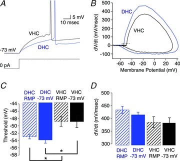
A, voltage responses to a somatic step current injection from −73 mV that yielded an action potential at approximately 50 ms for a DHC (blue) and VHC (black) neuron. B, phase plane plot for the representative action potentials in A. The dashed line represents 20 mV ms−1. C, threshold was significantly more depolarized in VHC neurones than DHC neurones when measured either from resting membrane potential (RMP; DHC n= 9, VHC n= 10; Wilcoxon RS test, P < 0.05) or −73 mV (DHC n= 8, VHC n= 9; Wilcoxon RS test, P < 0.05). D, the maximum dV/dt was not significantly different between DHC and VHC neurones when measured from RMP (DHC n= 9, VHC n= 10; Student's t test, P > 0.05) or from −73 mV (DHC n= 8, VHC n= 9, Student's t test, P > 0.05).