
Figure 1. The regions of subunits studied.
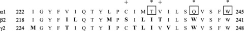
The figure shows the aligned TM1 regions, which were the regions swapped from the α1 subunit to the other subunits. All residues shown for the α1 sequence were directly substituted for the residues shown for the β2 or γ2L sequence. The critical residues in the α1 subunit are boxed and marked with asterisks (α1(T236), α1(Q241) and α1(W245)). Aligned residues that differ from the sequence in the α1 subunit are shown in bold type. The sequences shown begin at α1(I222), β2(I218) and γ2L(M224) (numbering for the mature subunit).