
Figure 7. GlyT1 are localized primarily on astrocytic processes.
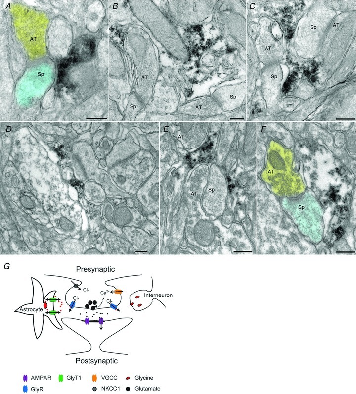
A, axon terminal (shaded yellow) makes synaptic contact with a dendritic spine (shaded blue). Electron-dense DAB reaction product coding for GlyT1 is visible within an astrocytic profile lying adjacent to the spine. B, an irregular astrocytic profile immunopositive for GlyT1 lies adjacent to two asymmetric synapses in the same electron microscopic field. The astrocytic cytoplasm has been disrupted by the immune processing and the relatively weak fixation necessary to preserve antigenicity. C, electron-dense reaction product in astrocytic profile lies between two axodendritic synapses. The small lightly-immunostained profile on the bottom left is likely also to be astrocytic. D, low-magnification montage shows immunostained astrocytic profile in a field of neuropil. E, axon terminal and spine are enveloped by immunostained astrocytic processes. F, axospinous synapse lies adjacent to immunopositive astrocytic process; false-colour as in A. G, illustration depicting the mechanism of preGlyR facilitation of neurotransmitter release. Abbreviations: AMPAR, AMPA receptor; AT, axon terminal; GlyR, glycine receptor; GlyT, glycine transporter; NKCC1, Na+–K+–2Cl− cotransporter; Sp, spine; VGCC, voltage-gated calcium channel. Scale bars: 200 nm.