
Figure 10. Density of active neurons in various brain regions controlling autonomic nervous activity.
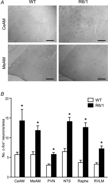
A, images of brain sections showing active neurons (black dots) detected by immunohistochemistry for c-Fos. Photomicrographs of coronal sections through the central (CeAM) and medial (MeAM) nuclei of the amygdala in 5.5-month-old wild-type (WT) and R6/1 mouse brains. Scale bars: 100 μm. B, bar graph showing grouped data of active neurons in brain regions that are known autonomic nervous centres (n= 4/group). CeAM and MeAM, central and medial nuclei of the amygdala, respectively; PVN, paraventricular nucleus of the hypothalamus; NTS, nucleus tractus solitarii; Raphe, raphe pallidus; RVLM, rostral ventrolateral medulla. *P < 0.05 vs. WT (ANOVA).