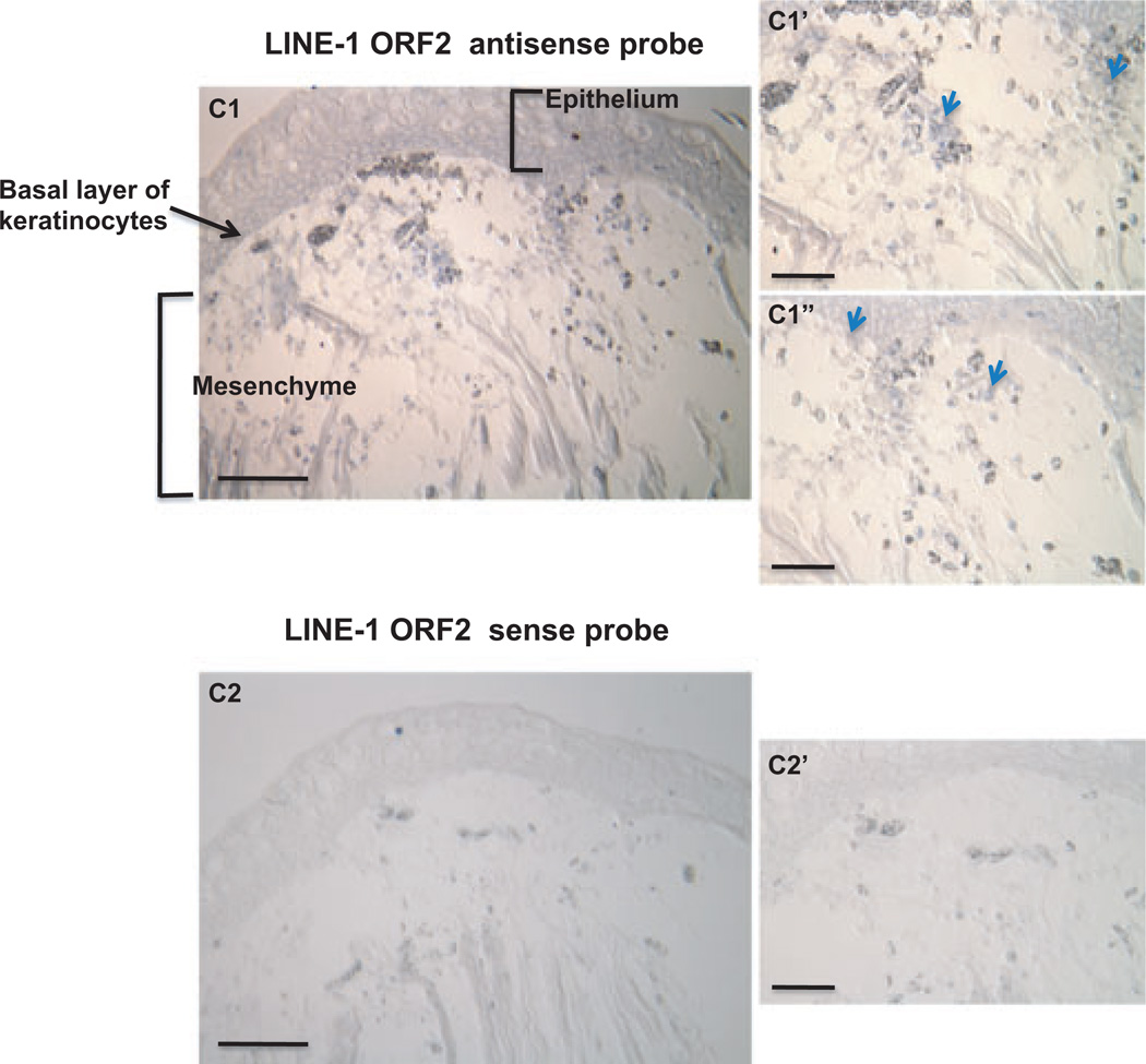
Fig. 3.
In situ hybridization of LINE-1 ORF-2 in 5 days post-amputation (pa) regenerating forelimb blastema. Sections were hybridized with either sense or anti-sense LINE-1 ORF2 RNA probe. The ORF2 sense RNA probe was used as control if the expression levels of the sense and the anti-sense transcripts were different. Staining (blue) was developed with 5-bromo-4-chloro-3-indolyl-phosphate (BCIP) and 4-nitroblue tetrazolium chloride (NBT) in alkaline phosphatase buffer after in situ hybridization (ISH) and anti-digoxygenin (DIG) alkaline phosphatase antibody incubation. Some of the cells bearing positive L1 signal were labeled by blue arrows. The scale bar for images C1 and C2 is 500 µm. The bar scale for images C1′, C1″, C2′ and C2″ is 200 µm. C1′, C1″ are the enlarged photos from different portions of the limb blastema presented in C1 similar to the relationship between C2′ and C2.

