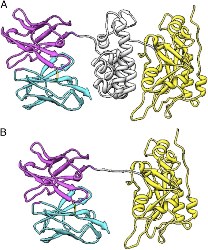
Fig. 1.
Structural model of RIT variants. (A) Structural model of Moxetumomab Pasudotox. The VL is in cyan, and the VH is in magenta. Domain II of the toxin is in gray, and domain III is in yellow. (B) HA22-LR. The linker containing the furin cleavage sequences is in gray. Image courtesy of Byungkook Lee (National Cancer Institute, NIH, Bethesda, MD).

