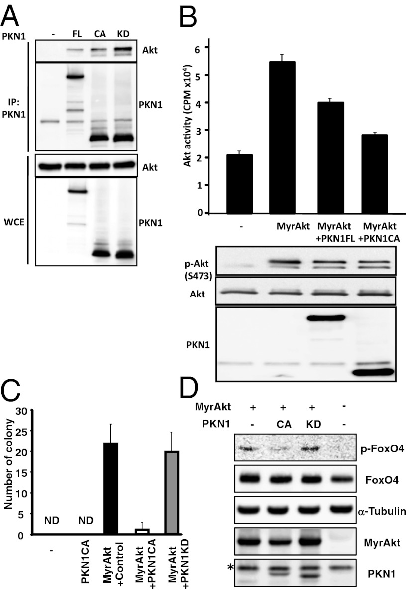
Fig. 2.
PKN1 interacts with and inhibits Akt. (A) 293T cells were transfected with expression vectors for HA-tagged Akt1, FLAG-tagged full-length (FL), and a constitutively activate (CA) or kinase-dead (KD) PKN1. Cell lysates were immunoprecipitated with anti-FLAG antibody-conjugated agarose and then immunoblotted with an anti-HA antibody or anti-FLAG antibody. (B) Akt activity was assessed using an in vitro Akt kinase assay that measured the phosphorylation levels of the Akt SGK substrate peptide. Data are representative of several different experiments. (C) Immortalized rat fibroblast 3Y1 cells were retrovirally transduced with myristoylated Akt1 (MyrAkt) together with GFP. Then, the MyrAkt (GFP)-positive cells were superinfected with retrovirus carrying PKN1CA or PKN1KD and plated in soft agar. Three weeks after plating, the number of colonies was determined. ND, not detected. (D) FoxO4 phosphorylation by MyrAkt in 3Y1 cells from C. *, nonspecific bands.

