Fig. 3.
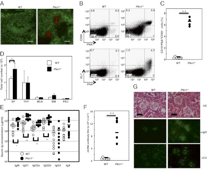
Immunological abnormalities in Pkn1−/− mice. Spontaneous germinal center (GC) formation in Pkn1−/− mice. (A) Spleen sections from WT and Pkn1−/− mice over 30 wk of age were stained with either an anti-IgM antibody (green) or peanut agglutinin (red). Magnification 100×. (B) Flow cytometric analysis of GC B cells from splenocytes that were stained with CD19, PNA, GL7, and CD95. (C) Statistical analysis of PNAhiCD95+ B cells in splenic CD19+ B cells. **P < 0.01, based on Student’s t test. (D) Total cell numbers in the spleen (SP), thymus (THY), mesenteric lymph node (MLN), bone marrow (BM), and peritoneal cavity (PEC) from either WT or Pkn1−/− mice. Data are from five mice per group. *P < 0.05 based on Student’s t test. (E) The Ig levels in sera from either WT (open circle) or Pkn1−/− (filled circle) mice were examined by ELISA. The bar in the figure shows the mean of 10–11 mice per group. *P < 0.05 and **P < 0.01, based on Student’s t test. (F) Anti-dsDNA antibody production in 1-y-old WT (n = 6) and Pkn1−/− mice (n = 5). (G) Development of glomerulonephritis in Pkn1−/− mice. Haematoxylin−eosin staining (HE, Top) and immunofluorescence staining with anti-IgG antibodies (αIgG, Middle) and anti-C3 antibodies (αC3, Bottom) of kidney sections from 6-mo-old WT and Pkn1−/− mice.

