Fig. 7.
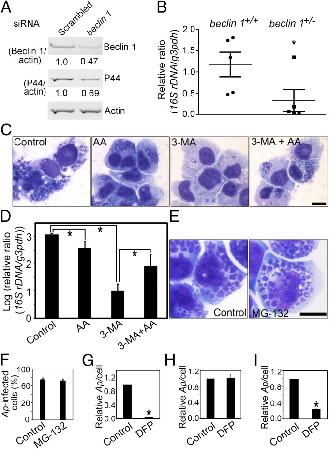
Beclin 1 depletion impairs Ap infection, and amino acid supplementation overrides 3-MA inhibition of Ap replication. (A) Immunoblotting of lysates of Ap-infected RF/6A cells transfected with beclin 1 siRNA or scrambled siRNA (2 d p.t., 3 d p.i.) using anti-P44, antiactin, and anti-Beclin 1. The values under the bands show the relative ratios of band intensities, with the ratios of those band intensities from scrambled siRNA set as one. (B) The dot plot of Ap load in the blood from beclin 1+/− and WT mice at 5 d p.i. Quantitative PCR of Ap 16S rDNA normalized to mouse G3PDH DNA. *Significantly different (P < 0.05). (C) Ap-infected cells: untreated (Control), or treated with amino acids supplementation (AA), 3-MA (3-MA), or 3-MA and amino acids (3-MA + AA). Diff-Quik staining. (Scale bar: 10 μm.) (D) Quantitative PCR of Ap 16S rDNA normalized to human G3PDH DNA. *Significantly different (P < 0.05) by ANOVA. (E and F) The effect of MG-132 on Ap growth. Diff-Quik–stained images of Ap in solvent control or MG-132–treated HL-60 cells (E) and the percentage of Ap infected HL-60 cells on 3 d p.i. (F). (Scale bar: 10 μm.) (G–I) The effect of DFP on Ap growth. Host cell-free Ap pretreated with solvent control or DFP was used to infect HL-60 cells (G). HL-60 cells were pretreated with solvent or DFP followed by washing and infection with Ap (H). HL-60 cells were infected with Ap followed by the treatment with solvent or DFP at 1 d p.i. (I). The relative ratios of Ap/cell were shown, with the ratios from control set as one. *Significantly different (P < 0.01).

