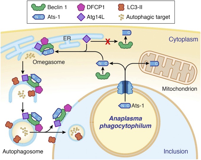
Fig. 8.
Proposed model of autophagosome nucleation by Ats-1 in Ap-infected cells. Ats-1 is translocated from Ap to the cytoplasm of infected cells by the type IV secretion system. A portion of Ats-1 interacts with the host autophagosome initiation complex (Atg14L-Beclin 1-Vps34), stimulating the formation of omegasomes in ER. Another portion of Ats-1 targets mitochondria, where it exerts antiapoptotic activity. The omegasome is marked with DFCP1. N terminus of Ats-1 is required for Atg14L recruiting and thus, autophagosome formation, but it is cleaved off when Ats-1 is imported into mitochondria. The isolation membrane elongates to envelop the cytoplasmic content into the double-membrane vacuole, the autophagosome, which is decorated with LC3. Ats-1 autophagosomes are recruited to Ap inclusion, and the outer membrane fuses with the Ap inclusion membrane, resulting in the release of the autophagic body-like content into the Ap inclusion.

