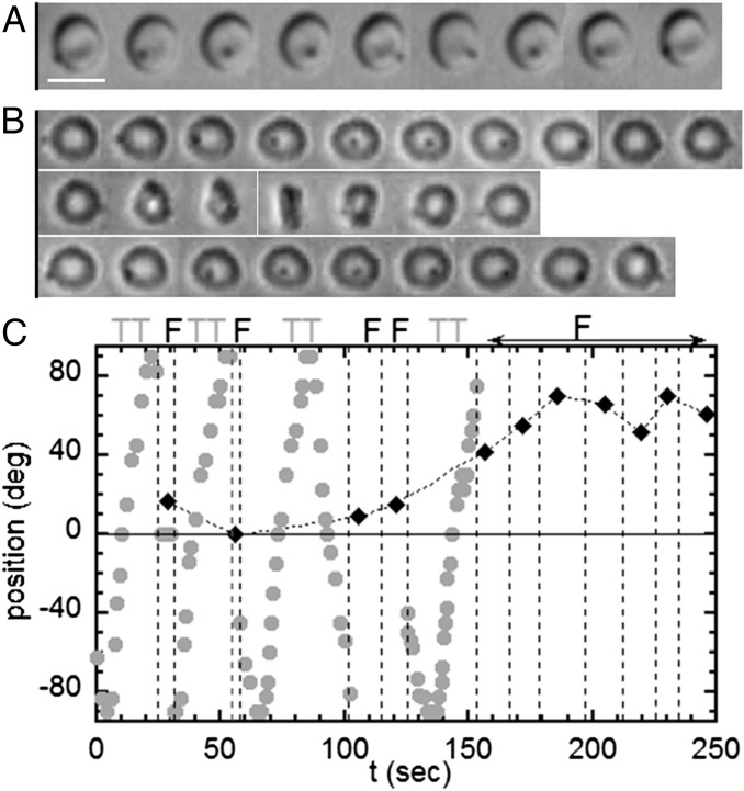
Fig. 4.
Tank-treading-to-flipping transition of RBCs whose membrane bears a latex bead (diameter, 1 μm); dextran 2 106 g/mol, c = 9% (wt/wt); scale bar, 8 μm; top-view observation. (A) Tank-treading RBC with rotation of the bead. The dimple of the biconcave shape is preserved (DIC image),  , time sequence of 5.48 s. (B) Intermittency at the transition. Sequences show TT, F, and TT, respectively;
  ; time sequence of 47.84 s. Tank-treading is detected from the rotation of the bead. The cell is still biconcave with the presence of the dimple (phase contrast image). (C) Transient intermittency. Dotted lines separate tank-treading from flipping regimes. ●, Variation of the bead position between −90° (1st image) to +90° (10th image) vs. time during the tank-treading–flipping transition at
  . ♦, Values of the angle φ during the flipping; when φ reaches 40°, the flipping regime is stabilized.

