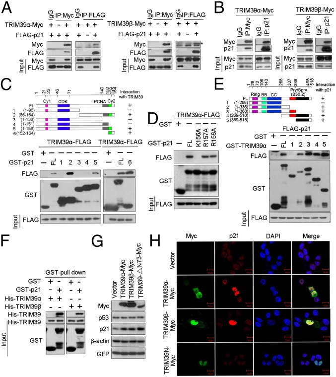
Fig. 2.
TRIM39 interacts with p21. (A) HEK293T cells were transiently transfected with plasmid DNA expressing Flag-p21 and/or TRIM39-Myc. Total cell lysates were immunoprecipitated with the indicated antibodies. The asterisk indicates the specific TRIM39β-Myc band. IP, immunoprecipitate. (B) HCT116 WT cells infected with the indicated lentiviral constructs were collected, and the cell lysates were immunoprecipitated with anti-Myc and anti-p21 antibodies. The immunoprecipitates were subjected to Western blotting with the indicated antibodies. (C and D) Extracts from HEK293T cells transfected with TRIM39α-FLAG were incubated with recombinant full-length (FL) GST-p21 or GST-p21 mutant coupled to GSH-Sepharose. Proteins retained on Sepharose were then blotted with the indicated antibodies. (E) Extracts from HEK293T cells transfected with FLAG-p21 were incubated with recombinant full-length GST-TRIM39α or GST-TRIM39α deletion mutant protein coupled to GSH-Sepharose. Proteins retained on Sepharose were then blotted with the indicated antibodies. (F) Purified His-TRIM39 proteins were incubated with GST-p21. Proteins retained on Sepharose were then blotted with the indicated antibodies. (G) HCT116 WT cells were transfected with the indicated expression constructs for 48 h. Cell lysates were then harvested and analyzed by Western blotting with the indicated antibodies. (H) HCT116 WT cells were transiently transfected with the indicated constructs for 48 h. Cells were then fixed and stained as indicated. (Scale bars, 10 μm.)

