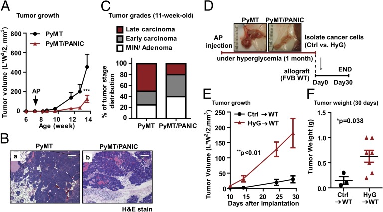
Fig. 1.
Hyperglycemic memory in mammary cancer cells result in the malignant progression. (A) Tumor growth for PyMT and PyMT/PANIC mice. Hyperglycemia was induced by dimerizer injection (AP20187; 0.5 μg/g per day for 5 d by i.p administration) in 7-wk-old PyMT/PANIC mice. Tumor growth was assessed by caliper measurements twice a week. n = 6 per group. ***P < 0.001 vs. PyMT by two-way ANOVA. (B) Representative images of H&E stain for PyMT and PyMT/PANIC. (Scale bars: 200 μm.) (C) Tumor grading was determined by using H&E-stained slides for 11-wk-old PyMT and PyMT/PANIC mice. n = 10 per group. (D) Schematic for the implantation of cancer cells taken from PyMT and PyMT/PANIC into isogenic wild-type hosts. (E and F) Tumor growth for hyperglycemia experienced cancer cells (HyG)-bearing mice compared with control cells (Ctrl)-bearing mice. Either HyG or Ctrl cancer cells (0.5 × 10⁶ per mouse) were implanted into the mammary adipose tissues of wild-type mice and monitored tumor growth by caliper measurement (E). Data represent mean ± SEM (n = 5 per group). **P < 0.01 vs. Ctrl by two-way ANOVA. Tumor weights (F) were determined at 30 d after implantation. *P = 0.038 vs. Ctrl by unpaired Student’s t test.

